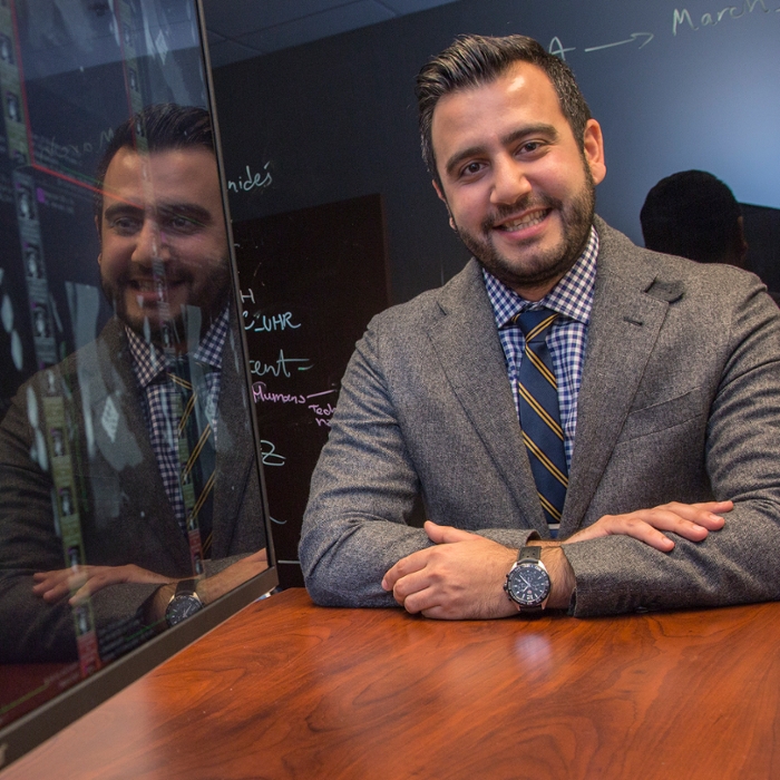
Amir Pourmorteza, PhD
Jan. 31, 2018

Amir Pourmorteza is a biomedical engineer by training, an artist by design, and an innovator by nature, which makes imaging sciences the perfect fit.
“I like to build things,” the assistant professor says simply. “As an engineer, I want to come up with an idea, create a prototype, and then go from bench to bedside, that is refine the innovation so it ultimately improves the way we care for patients. Imaging is especially interesting because it’s visual. It has an artistic edge to it. You can see what you’re doing and see in pictures the results of your innovations.”
Dr. Pourmorteza is especially focused on spectral and high-resolution CT, imaging technologies with the potential to transform the way we see and understand processes in the body. Until very recently, Pourmorteza explains, all CT scanners were analog and imaged in black and white, with plenty of shades of gray. Spectral (photon-counting) CT employs a high-resolution digital spectral detector to more precisely and fully map organs and visualize abnormalities while simultaneously limiting radiation dosage exposure. It is works as if we can see the different colors of the x-ray spectrum.
“Contrast dye only highlights something but doesn’t differentiate what it is,” he explains. “Currently, we inject patients with a contrast dye and then take a series of CT images over time to see how and where the dye moved through the organ in order to detect tumors or other abnormalities. This gives the radiologist a series of images taken over time to interpret. Spectral imaging would allow us to inject differently-hued contrast dyes at different times and then image once to see how and where the dyes moved through the organ, which exposes the patient to radiation only once. The radiologist then would have a visual map showing all these color-coded, timed doses of contrast on a single image.”
As part of his work, Pourmorteza will have to develop protocols for calculating the contrast amounts and timing of injections for various organ studies, work the engineer in him relishes. The scientist then will further develop clinical workflows and visualization schemes to maximize the power of color CT imaging, the element of artistry in his work.
Pourmorteza also is working on harnessing the power of high-resolution CT imaging to help radiologists visualize smaller and smaller pieces in the body. With higher resolution imaging, he hopes to empower radiologists, for example, to find tumors earlier in their development, to see coronary plaques in their earliest stages of accumulation, and even to see smaller blood vessels to catch nascent disease activation.
Another aspect of his high-resolution research includes quantification of heart muscle contractility. This work builds on advances he made in cardiac imaging for his doctoral dissertation at Johns Hopkins University, and his tenure at the National Institutes of Health where he led the photon-counting group. At Emory, he’s collaborating with a company to bring the cardiovascular innovations to market. At the same time, he’s at work with Arthur Stillman, MD, William and Kay Cassarella Professor of Radiology and Imaging Sciences and director of cardiothoracic imaging, and Ernest V. Garcia, PhD, endowed professor and director of the Emory Nuclear Cardiology Research and Development Lab, on developing algorithms to extract and better use the full range of information captured in cardiac CT.
Along those same lines, Pourmorteza will be working on ways to better harness the power of artificial intelligence or machine learning in collaboration with Shamim Nemati, PhD, assistant professor in the Department of Biomedical Informatics. “With AI, it’s a situation of garbage in/garbage out. If we don’t have precise and accurate ways to generate good data in the first place, our AI algorithms will produce useless information. My focus is not on developing new AI models, it’s on generating good data to train these models.”
Pourmorteza also is collaborating with Phuong-Anh Duong, MD, associate professor and director of CT imaging for Emory Healthcare and Dr. Rahnliang Hu, MD, assistant professor and neuroradiologist, to leverage AI for quality control particularly for neuro and cardiac imaging.
Pourmorteza was drawn to Emory because of its eminence in biomedical sciences as well as its reputation for innovation. He also was drawn to what seemed to be at atmosphere of collegiality and collaboration. Now that he’s been at Emory for several months, he’s even more impressed by the depth and breadth of such collaboration, something he finds uncommon in top research universities.
“Of course, the partnership between GA Tech and Emory was a huge draw for me,” he says. “The culture of collaboration is very exciting. People are very open and eager to collaborate, not just within the department, but across the university and across institutions. This cross-pollination, is so rewarding and different from the siloed approach seen elsewhere.”
That both Emory Radiology’s chair, Carolyn Meltzer, MD, and Emory’s executive vice president for health affairs and chief executive officer of Emory Healthcare, Jonathan Lewin, MD, also hail from Johns Hopkins delights Dr. Pourmorteza.
“Clearly, Emory is the place for me to be.”


